In the rapidly evolving landscape of medical diagnostics, staying ahead of technological advancements is not merely an advantage—it’s a necessity. For R&D engineers and professionals in the medical imaging sector, the latest software releases from pioneers like QT Imaging represent critical inflection points. The urgency to understand and integrate these innovations is paramount, as they directly impact diagnostic accuracy, clinical workflow efficiency, and ultimately, patient outcomes. Today, we delve into the significant implications of QT Imaging’s latest offering: the next generation of breast imaging software, version 4.5.0.
Unveiling QT Imaging’s Next Generation Breast Imaging Software (v4.5.0)
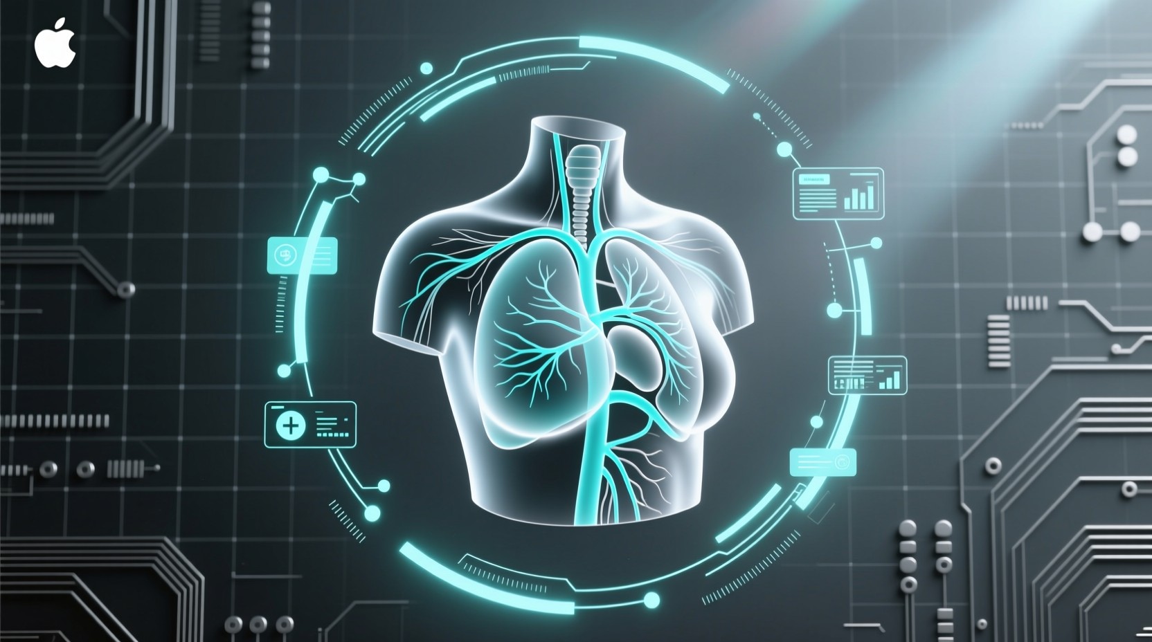
QT Imaging has officially launched version 4.5.0 of its image reconstruction software for its QTI Breast Acoustic CT system. This release signifies a substantial leap forward in breast imaging technology, building upon the company’s commitment to providing radiation-free, high-resolution, and comprehensive breast health solutions. The update is not just an incremental improvement; it introduces a suite of advanced features designed to enhance image quality, deepen tissue characterization, and broaden clinical applicability.
Deep Technical Analysis: What’s New in v4.5.0?
At the core of version 4.5.0 are several key technical enhancements:
- Improved Spatial Resolution in Reflection Imaging: The software now incorporates optimized, spatially varying deconvolution techniques during image reconstruction. This algorithmic refinement leads to more accurate image representation, a critical factor in identifying subtle abnormalities. The precise nature of this deconvolution process, while proprietary, is understood to involve advanced signal processing to minimize artifacts and enhance detail, directly addressing the challenge of achieving high fidelity in complex tissue structures.
- Enhanced Reflection Images via Data Fusion: A groundbreaking feature in this release is the introduction of enhanced reflection images generated by fusing speed-of-sound (SoS) and reflection data. This multimodal approach provides clinicians with richer visualization and more comprehensive tissue characterization information than previously possible with reflection imaging alone. The fusion algorithm intelligently combines the acoustic properties derived from SoS measurements with the signal intensity from reflection data, offering a more nuanced understanding of tissue composition and potential pathology. This is a significant architectural decision, moving beyond single-modality analysis to a more integrated, data-driven approach.
- Optimized Reconstruction for Specific Anatomies: Version 4.5.0 includes improved image reconstruction capabilities specifically for smaller breasts. This addresses a common challenge in breast imaging, ensuring that diagnostic accuracy is maintained across a wider range of patient anatomies. Furthermore, the update introduces more accurate assessment of fibroglandular tissue composition in breasts with implants. This enhancement is crucial for differentiating between implant material, surrounding tissue, and potential pathologies, often a complex task with conventional imaging methods.
- Development of Attenuation as a Quantitative Biomarker: QT Imaging is actively developing attenuation as an additional quantitative biomarker. This is expected to complement existing speed-of-sound and reflection intensity measurements, providing clinicians and researchers with even more quantitative data for improved analysis and decision support. The integration of attenuation, a measure of how sound waves lose energy as they pass through tissue, promises to further refine tissue characterization capabilities.
While specific benchmark numbers for v4.5.0 are not publicly detailed, the previous version (4.4.0) leveraged NVIDIA’s L40 GPU acceleration, achieving substantial reductions in image processing time. This suggests that v4.5.0 likely maintains or improves upon these performance gains, ensuring efficient workflows even with more complex reconstructions and data fusion.
Background Context: QT Imaging’s Vision for Breast Health
QT Imaging is dedicated to transforming breast health management through innovative, radiation-free imaging technology. Their QTI Breast Acoustic CT system is a non-invasive technology that creates 3D breast images without compression, contrast agents, or ionizing radiation. This core technology, based on low-frequency sound waves, has consistently aimed to provide a safer, more comfortable, and more informative alternative to traditional mammography and other breast imaging modalities. The platform approach, integrating AI, advanced analytics, and cloud connectivity, underscores QT Imaging’s commitment to a scalable and evolving ecosystem for breast imaging.
Practical Implications for R&D and Clinical Teams
The release of version 4.5.0 carries significant practical implications for various stakeholders:
- For R&D Engineers: This update offers new avenues for research and development. The enhanced data fusion techniques and the ongoing development of attenuation as a biomarker present opportunities for exploring novel algorithms, improving diagnostic models, and pushing the boundaries of quantitative ultrasound imaging. Understanding the underlying deconvolution algorithms and data fusion methodologies will be key to leveraging these advancements.
- For Radiologists and Clinicians: The improved spatial resolution and enhanced tissue characterization directly translate to better image interpretability and potentially higher diagnostic accuracy. The specific improvements for small breasts and breasts with implants address known clinical challenges, expanding the utility of the QTI system across a broader patient population. This can lead to more confident diagnoses and more personalized treatment plans.
- For Infrastructure and IT Teams: While specific security patches or CVE IDs are not detailed in the release notes, any software update in the medical field necessitates a review of security protocols. Ensuring data integrity, patient privacy, and compliance with regulations like HIPAA remains paramount. The integration of new data types (e.g., fused SoS and reflection data) may also require adjustments to storage and processing infrastructure.
Security and Migration Considerations
As with any significant software update, particularly in the sensitive domain of medical imaging, security and migration are critical aspects. QT Imaging has not publicly disclosed any specific CVE IDs or critical security patches associated with version 4.5.0. However, it is standard practice for medical device software updates to undergo rigorous validation and security testing. Organizations implementing this update should:
- Review Release Notes Thoroughly: While detailed changelogs are not always publicly available, R&D and IT teams should seek out any documentation provided by QT Imaging regarding the update, including any security-related advisories or best practices.
- Perform Rigorous Testing: Before full deployment in a clinical setting, thorough testing in a non-production environment is essential. This includes functional testing, performance benchmarking, and integration testing with existing systems.
- Ensure Data Compatibility: Verify that the new version maintains backward compatibility with existing patient data and integrates seamlessly with PACS (Picture Archiving and Communication System) and other relevant healthcare IT infrastructure.
- Maintain Audit Trails: Implement robust logging and auditing mechanisms to track software usage, any configuration changes, and system performance, which is crucial for regulatory compliance and troubleshooting.
Best Practices for Adoption and Integration
To maximize the benefits of QT Imaging’s next-generation software, consider the following best practices:
- Invest in Training: Ensure that all clinical and technical staff are adequately trained on the new features and functionalities of version 4.5.0. Understanding the nuances of the enhanced reflection images and improved tissue characterization is vital for effective utilization.
- Collaborate with QT Imaging Support: Engage with QT Imaging’s technical support and clinical specialists to facilitate a smooth integration process and to address any specific implementation challenges.
- Monitor Performance Metrics: Continuously monitor key performance indicators (KPIs) related to image quality, processing times, diagnostic accuracy, and user feedback. This data will be invaluable for assessing the impact of the update and identifying areas for further optimization.
- Stay Informed on Future Developments: QT Imaging is actively developing attenuation as a biomarker. R&D teams should stay abreast of these developments, as they represent the next frontier in quantitative breast imaging.
Related Internal Topics
- Advanced Ultrasound Imaging Techniques
- AI in Medical Diagnostics: Trends and Applications
- Medical Imaging Software Development Best Practices
Conclusion: A Forward-Looking Perspective
The release of QT Imaging’s version 4.5.0 breast imaging software marks a significant advancement in the field, offering enhanced resolution, sophisticated data fusion for superior tissue characterization, and improved utility across diverse patient anatomies. For R&D engineers, this update presents exciting opportunities for innovation in algorithm development and quantitative biomarker exploration. For clinical teams, it promises greater diagnostic confidence and workflow efficiency. As QT Imaging continues to push the envelope with innovations like the development of attenuation biomarkers, the future of breast imaging is increasingly multimodal, data-rich, and non-invasive. Embracing these technological leaps is essential for any organization committed to providing state-of-the-art breast health management.